
Our lab develops highly specialized instrumentation for generating the intense acoustic fields required for histotripsy research. This includes transducers that convert electrical energy into sound and electrical drivers that control timing of acoustic bursts and capture acoustic echoes from tissue and cavitation.
Rapid Prototyping Ultrasound Transducers
High power ultrasound transducers are key to generating the microsecond-length, very high pressure ultrasound pulses required for histotripsy. Our lab has been using rapid prototyping (PR) methods for more than ten years to construct ultrasound transducers [1,2], allowing for fast design iteration at low cost customized to particular applications and experiments. The transducers we have constructed span the range from small endoscopic probes [3] to 30 centimeter diameter hemispheres with hundreds of module elements for non-invasive histotripsy treatments in the brain [4]. The design process starts with simulations of the desired acoustic output from which we form a CAD model (solidworks or inventor). We have characterized the acoustic properties of a range of rapid prototyping materials allowing us to design acoustic lenses for focusing sound or quarter-wave matching layers for optimizing output of phased-arrays. We purchase and stock larger flat pieces of piezoelectric materials which we then cut by water jet or dicing saw as needed for particular designs. These are then bonded to the housings, lenses, and/or matching layers constructed from rapid prototyping to form complete transducers. The versatility and lower fabrication costs afforded by RP methods may be beneficial in the development of complex transducer geometries suitable for a variety of research and clinical applications.
Phased Arrays
We have used our rapid prototyping method to construct large phased array transducers. For example, a 750-kHz truncated circular aperture array (165 mm x234 mm) transducer with a focal length of 142 mm was designed and built for liver cancer treatment (Fig. 1A)[2]. The aperture was segmented into 260 arc-shaped modular elements, each approximately 11.5 mm x11.5 mm, arranged in concentric rings. The resulting aperture utilization was 92%. The full-width-half-maximum (FWHM) focal zone of the array was measured to be 1.6 mm x1.1 mm x4.5 mm, and the FWHM electrical steering range was measured to be 38.5 mm x33 mm 40 mm. The array was estimated to be capable of generating approximately 120-MPa peak negative pressure at the geometric focus. Another example is the 700kHz, 360-element, 30-cm diameter hemispherical transcranial histotripsy array transducer. The histotripsy array (Fig. 1B) generates an estimated peak negative pressure up to 300MPa and a focal zone of 1.2×1.2×2.3 mm in free field. The -6dB electronic focal steering range is 33 mm laterally and 50 mm axially.
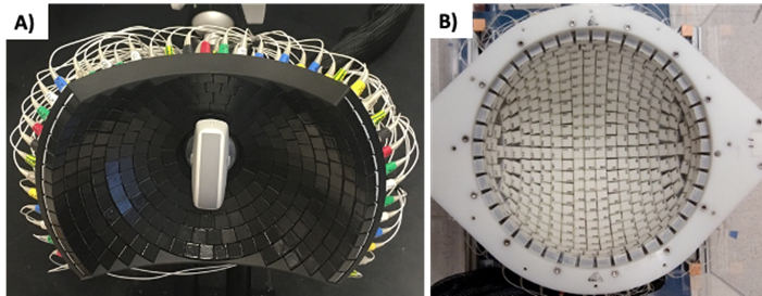
Fig. 1: A) 750kHz, 260-element liver histotripsy array and B) 700kHz, 360-element transcranial histotripsy array.
Electrical Driving Systems
Piezoelectric transducers for generating histotripsy must be excited by short electrical tone bursts of thousands of volts and up to thousands of amps. The timing of the bursts need to be adjustable up to tens of microseconds with a precision of tens of nanoseconds for phased-array electronic steering or aberration correction. In addition, the latest histotripsy array transducers must switch rapidly within microseconds to receiving small acoustic echoes from tissue and emissions from cavitation with high fidelity for treatment monitoring and aberration correction. This work is a challenging combination of novel high voltage electrical circuits with precision analog to digital conversion and high throughput digital electronics and embedded controllers. We use the latest SiC transistors and diodes for high power efficiency on transmit and AFEs designed for ultrasound imaging on receive. Control and data streaming is performed by a set of networked system on a chip FPGAs with dual core ARM processors running linux. We design all electrical circuitry and software in-house and we maintain a prototyping space for microelectronics assembly and testing new designs.
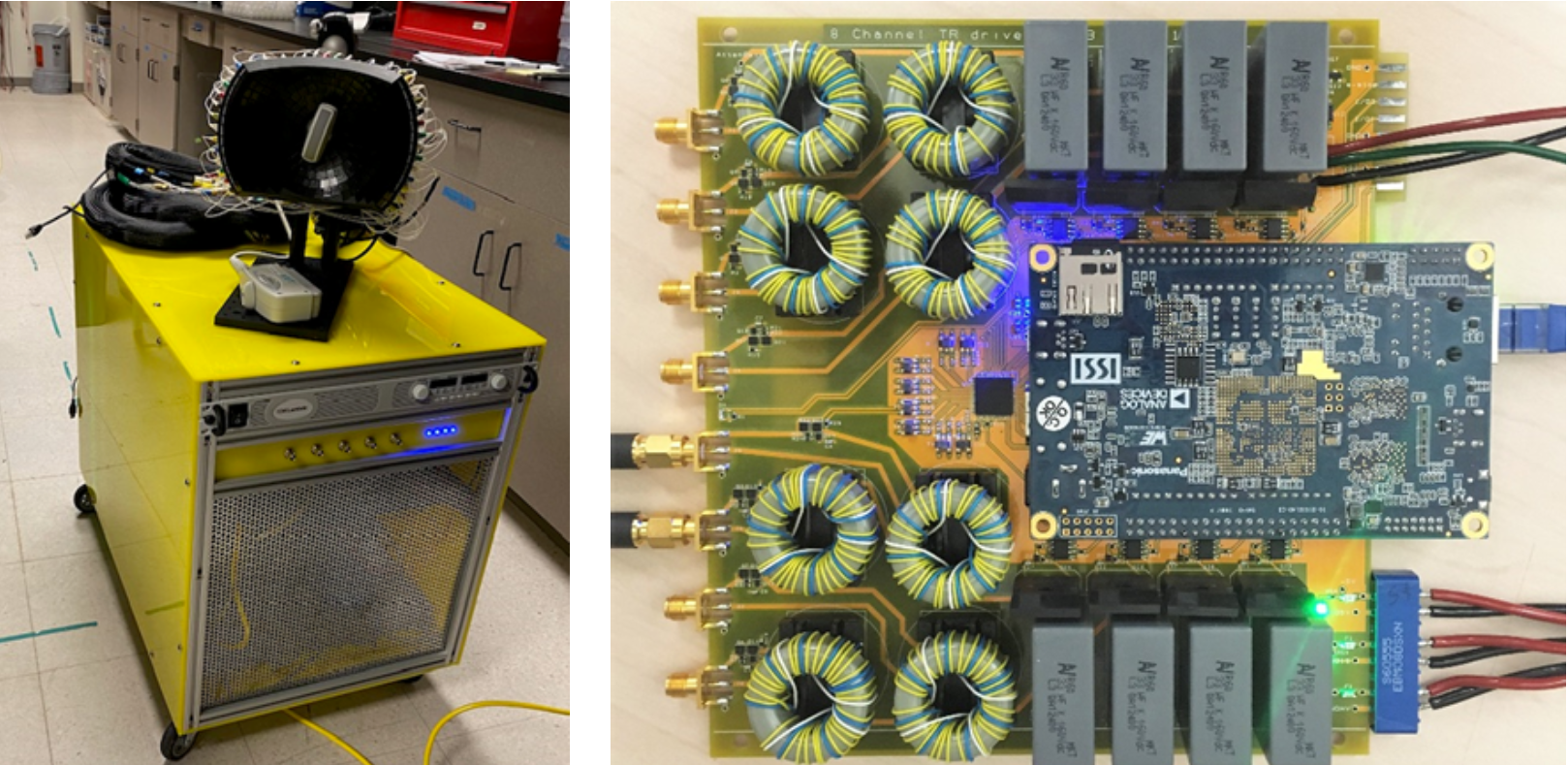
Fig. 2: Left) 260 channel phased array over transmit-receive driving system unit. Right) An eight channel circuit board from the system.
References
[1] Kim, Y., Maxwell, A. D., Hall, T. L., Xu, Z., Lin, K. W. & Cain, C. A. Rapid prototyping fabrication of focused ultrasound transducers. IEEE Trans Ultrason Ferroelectr Freq Control 61, 1559-1574, (2014).
[2] Stocker, G. E., Lundt, J. E., Sukovich, J. R., Miller, R. M., Duryea, A. P., Hall, T. L. & Xu, Z. A Modular, Kerf-Minimizing Approach for Therapeutic Ultrasound Phased Array Construction. IEEE Trans Ultrason Ferroelectr Freq Control 69, 2766-2775, (2022). PMC9594968
[3] Stocker, G. E., Zhang, M., Xu, Z. & Hall, T. L. Endocavity Histotripsy for Efficient Tissue Ablation- Transducer Design and Characterization. IEEE Trans Ultrason Ferroelectr Freq Control 68, 2896 – 2905, (2021).
[4] Lu, N., Hall, T. L., Choi, D., Gupta, D., Daou, B. J., Sukovich, J. R., Fox, A., Gerhardson, T. I., Pandey, A. S., Noll, D. C. & Xu, Z. Transcranial MR-Guided Histotripsy System. IEEE Trans Ultrason Ferroelectr Freq Control 68, 2917-2929, (2021). PMC8428576